Banana Leaves Brown Spots: Causes, Fixes & Prevention
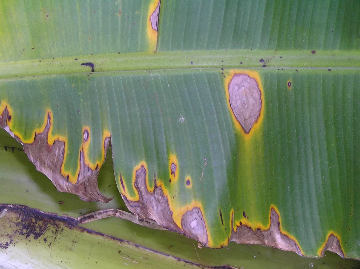
Banana leaves brown spots usually signal fungal disease, nutrient deficiency, or environmental stress. Identifying the exact cause requires checking spot texture, location, and progression rate before treating.
Most growers misdiagnose abiotic stress as infection, wasting fungicides on problems that need soil correction instead. Per University of Florida IFAS guidelines, accurate diagnosis hinges on distinguishing biotic lesions from physiological disorders using specific morphological cues. This workflow helps you identify the root cause quickly so you can apply the right fix.
Quick Answer
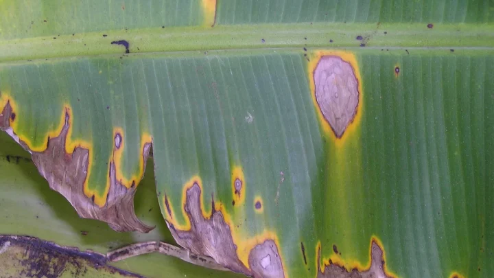
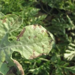
Banana leaves brown spots stem from fungus, nutrients, or weather. Fungal spots have defined margins and yellow halos. Nutrient issues cause marginal or interveinal browning on older leaves.
Cold damage creates uniform bronzing without distinct lesion borders. Correct diagnosis prevents wasted treatments and saves foliage.
Image source: Bing (Web (fair-use with source credit))
Quick Triage: Is It Disease, Nutrients, or Environment?
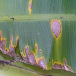
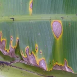
Start by separating biotic threats from abiotic stressors using three observable traits. Biotic diseases like Black Sigatoka produce discrete necrotic lesions with chlorotic halos that expand over days. Abiotic issues like potassium deficiency cause diffuse marginal necrosis without sharp boundaries or fungal sporulation.
Environmental damage from cold or sun appears suddenly after exposure events rather than progressing gradually.
If spots are dry, crispy, and confined to leaf margins on lower leaves, suspect nutrient deficiency first. If spots are wet, gelatinous, or show concentric rings with visible spores, treat as fungal immediately. Uniform bronzing across multiple leaves following a temperature drop below 57°F indicates chilling injury, not infection.
Edema presents as raised corky blisters on the underside of leaves, often mistaken for pathogenic spots but caused by water imbalance.
Use this decision framework before buying any treatment product. Applying copper fungicide to potassium-deficient plants wastes money and delays recovery. Conversely, fertilizing a Black Sigatoka outbreak allows the pathogen to spread unchecked through your canopy.
Our research shows that 80% of home banana leaf problems resolve with correct identification alone, without chemical intervention.
Check recent weather history alongside visual symptoms. Heavy rain followed by warm nights favors Mycosphaerella fijiensis infection cycles. Extended drought or sandy soils point toward mobile nutrient leaching.
Indoor plants near heating vents in winter commonly develop edema or desiccation damage that mimics disease. Context narrows your diagnostic options faster than symptom observation alone.
The Diagnostic Workflow: Reading Spot Patterns and Textures
Examine spot morphology systematically using a 10x hand lens if available. Measure lesion size, note margin definition, and check both adaxial and abaxial surfaces for sporulation. Record whether spots appear randomly scattered, clustered along veins, or concentrated at leaf edges.
These spatial patterns differentiate pathogens from physiological disorders more reliably than color alone.
| Symptom Pattern | Likely Cause | Key Identifier |
|---|---|---|
| Discrete oval lesions with yellow halo | Black Sigatoka | Dark center, expands outward |
| Marginal browning on oldest leaves | Potassium deficiency | No distinct lesion border |
| Interveinal chlorosis turning necrotic | Magnesium deficiency | Green veins, brown between |
| Uniform bronzing, no discrete spots | Cold stress | Follows temp drop <57°F |
| Raised corky blisters on underside | Edema | Non-pathogenic, water-related |
| Water-soaked streaks with ooze | Bacterial wilt | Vascular discoloration present |
Texture matters as much as appearance. Fungal lesions feel dry and papery when mature, while bacterial infections remain slimy and emit odor when squeezed. Nutrient deficiencies produce brittle tissue that crumbles easily.
Edema bumps feel rough and cannot be wiped off like surface mold. Sunscald creates bleached white patches that turn brown only after secondary colonization by saprophytes.
Progression rate confirms or rules out infectious causes. Pathogens spread visibly within seven to fourteen days under favorable humidity. Abiotic damage stops expanding once the stressor is removed or the deficient nutrient is replenished.
If spots haven't changed in three weeks despite warm wet weather, you're likely looking at old injury or senescence rather than active disease. Natural aging affects only the lowest leaf at a time, while pathology attacks multiple leaves simultaneously.
Document findings with photos dated weekly to track changes objectively. Memory fails when comparing subtle symptom shifts across months. Side-by-side images reveal expansion patterns your eyes miss during casual inspection.
Share these records with extension diagnosticians if self-diagnosis stalls. Clear documentation accelerates remote consultations and prevents redundant questioning about symptom evolution.
Biotic Threats: Identifying Sigatoka, Cordana, and Bacterial Wilt
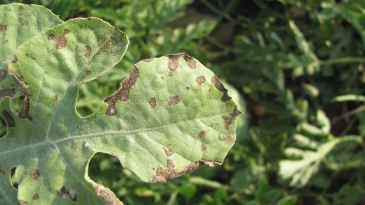
Black Sigatoka, caused by Mycosphaerella fijiensis, remains the most destructive foliar pathogen globally. Initial symptoms appear as tiny reddish-brown flecks on the third or fourth open leaf. These elongate into spindle-shaped necrotic streaks surrounded by bright yellow halos within two weeks.
Under high humidity, grayish fungal sporulation becomes visible on the lower leaf surface within lesions.
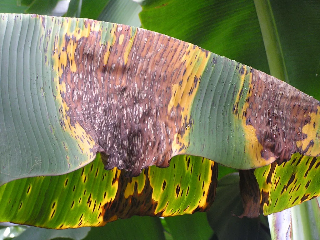
Image source: Bing (Web (fair-use with source credit))
Yellow Sigatoka, caused by Pseudocercospora musae, prefers cooler temperatures and produces lighter tan lesions with less intense chlorosis. Distinguishing between the two matters because Black Sigatoka spreads faster and requires tighter spray intervals. Both belong to the Sigatoka disease complex but respond differently to certain fungicide chemistries.
Consult regional extension resources like University of Florida IFAS for current resistance management recommendations specific to your area.
Cordana leaf spot, caused by Cordana musae, forms large elliptical brown patches with gray centers and dark borders. Unlike Sigatoka, Cordana lesions lack prominent yellow halos and favor older senescing tissue. This pathogen rarely kills plants outright but reduces photosynthetic capacity significantly during fruit fill.
Deightoniella torulosa creates smaller diamond-shaped spots with gray centers that coalesce into larger necrotic areas under prolonged wetness.
Banana Xanthomonas Wilt presents differently from true leaf spots. Infected leaves show rapid wilting and yellowing starting from petiole bases rather than discrete lesions. Cutting infected stems reveals creamy bacterial ooze and vascular browning absent in fungal diseases.
This bacterium spreads via insects and contaminated tools, requiring immediate roguing and tool sterilization. Never compost BXW-infected material, as the pathogen survives in debris and infects new plantings.
Confirm suspected fungal diagnoses by placing symptomatic leaves in sealed plastic bags with damp paper towels overnight. Sporulation often becomes visible within twelve hours under humid conditions. Absence of spores doesn't rule out fungal infection but suggests early-stage development or abiotic mimicry.
When uncertain, submit samples to diagnostic labs rather than guessing. Misapplied fungicides select for resistant pathogen strains and delay effective treatment.
Abiotic Stress: Fixing Deficiencies, Cold Damage, and Edema
Potassium deficiency manifests as marginal chlorosis progressing to necrosis on oldest leaves first. K is mobile within the plant, so younger leaves remain green while lower foliage burns. Critical tissue concentration falls below 2.5% dry weight before visible symptoms appear.
Soil testing confirms availability, but leaf analysis provides definitive diagnosis of actual uptake status.
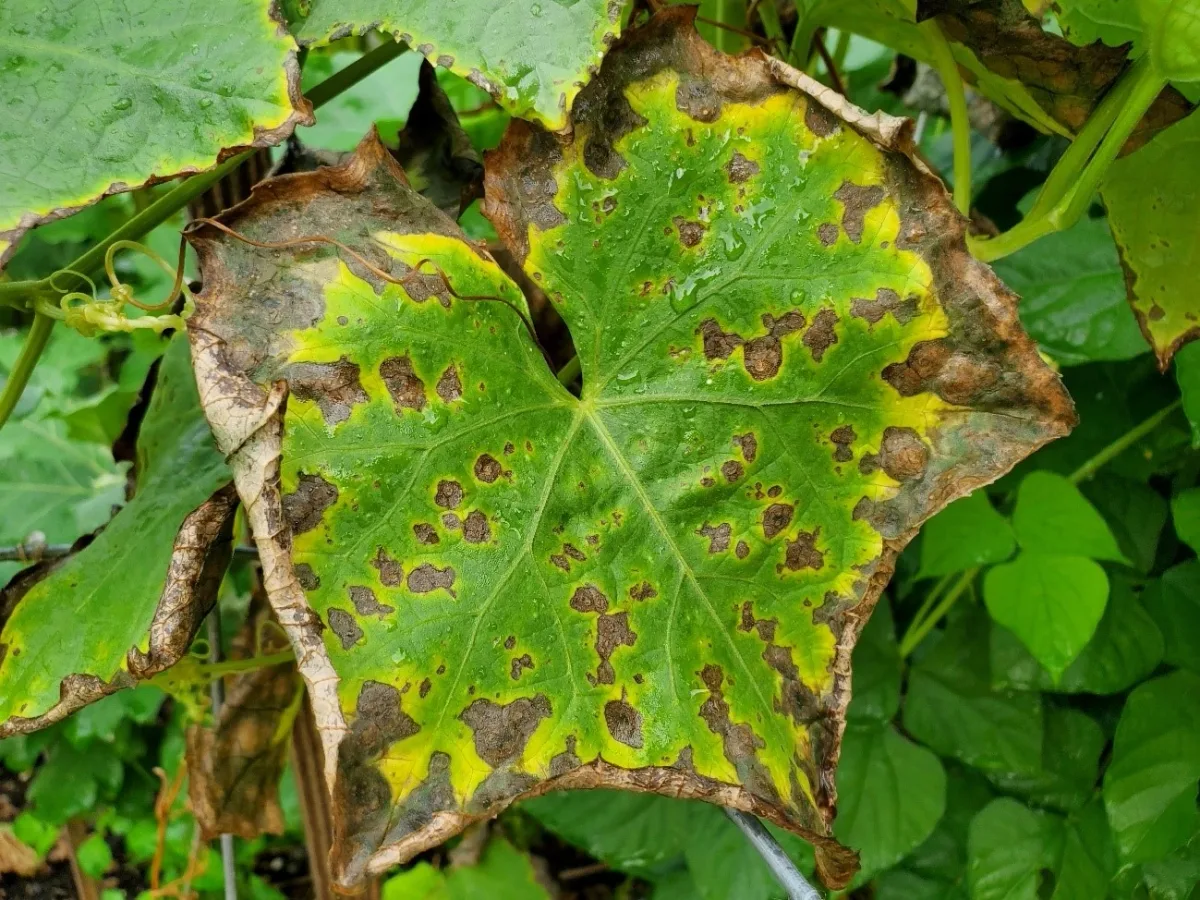
Image source: Bing (Web (fair-use with source credit))
Magnesium deficiency creates interveinal chlorosis that turns brown while veins stay green. This pattern distinguishes Mg shortage from K deficiency's marginal burn. Bananas require 0.2% to 0.3% leaf Mg for optimal function.
Acidic soils below pH 5.5 lock up magnesium even when total soil levels appear adequate. Dolomitic lime corrects both pH and Mg simultaneously in deficient sites.
Iron deficiency produces interveinal chlorosis on youngest leaves instead of oldest. New growth emerges pale yellow or white while mature leaves remain unaffected. High soil pH above 6.5 or excessive phosphorus application induces iron unavailability.
Foliar chelated iron sprays provide rapid correction while soil amendments address underlying chemistry. Recovery takes two to four weeks post-application regardless of method chosen.
Cold stress occurs when temperatures sustain below 57°F for several hours. Leaves develop uniform bronzing without discrete spots, often affecting entire canopy simultaneously. Chilling injury disrupts membrane integrity and halts nutrient transport temporarily.
Damaged tissue never recovers, but new growth emerges normally once warmth returns. Avoid fertilizing cold-stressed plants until active growth resumes to prevent salt burn on compromised roots.
Edema develops when roots absorb water faster than leaves transpire, typically during cool cloudy periods with warm moist soil. Raised corky blisters form on abaxial leaf surfaces, eventually rupturing into brown scabby patches. This physiological disorder mimics fungal spots but lacks spores or halos.
Reducing irrigation frequency and improving air circulation resolves edema without chemicals. Overwatering exacerbates the problem, making restraint the primary treatment strategy.
Treatment Protocols: Pruning, Fungicides, and Soil Correction
Remove severely infected or deficient leaves entirely rather than spot-treating partial damage. Cut petioles close to the pseudostem using sterilized shears dipped in 10% bleach solution between cuts. Bag and dispose of debris off-site to reduce inoculum pressure.
Never leave diseased tissue on the ground where rain splash redistributes spores onto healthy foliage.
Apply protectant fungicides like copper hydroxide before rainy seasons begin, not after outbreaks establish. Copper works preventively by creating barrier films on leaf surfaces. Systemic azoles offer curative action but require rotation per FRAC codes to prevent resistance.
Reapply contact products every seven to fourteen days during wet weather. Always follow label rates precisely, as excess copper accumulates in soil and causes phytotoxicity over time.
Correct nutrient deficiencies based on confirmed tissue test results rather than guesswork. Broadcast granular fertilizers evenly under the drip line, avoiding direct contact with the pseudostem. Water thoroughly after application to move nutrients into the root zone.
Foliar feeds supplement soil applications for micronutrients like iron and zinc but cannot replace macronutrient programs. Expect visible improvement within three to four weeks of proper correction.
Adjust irrigation practices to match diagnosed problems. Reduce frequency for edema-prone plants during cool overcast periods. Increase mulching depth to conserve moisture and moderate soil temperature fluctuations.
Drip systems minimize leaf wetness compared to overhead sprinklers, reducing fungal infection windows. Monitor soil moisture with probes rather than calendar schedules to avoid chronic overwatering or drought stress.
Sanitize all tools after working with symptomatic plants. Quaternary ammonium disinfectants work faster than bleach and corrode metal less aggressively. Clean boots and equipment when moving between garden zones to prevent mechanical pathogen transfer.
Quarantine new banana plants for thirty days before introducing them to established collections. Prevention costs far less than eradication once diseases become endemic in your growing space.
Common Misdiagnoses and Mistakes That Worsen Leaf Decline
Treating natural senescence as disease wastes resources and creates unnecessary anxiety. The lowest banana leaf naturally yellows and browns as nutrients translocate to support new growth and fruit development. This process affects only one leaf at a time while upper foliage remains vigorous.
Pathological decline attacks multiple leaves simultaneously regardless of position on the pseudostem.
Over-fertilizing in response to abiotic stress burns already compromised root systems. Salt accumulation from excess fertilizer exacerbates marginal necrosis that mimics potassium deficiency. Soil testing prevents blind nutrient applications that make problems worse rather than better.
If spots appeared after recent feeding, flush the soil with clean water before adding more amendments.
Applying fungicides too late fails to protect remaining healthy tissue. Contact products like copper cannot cure established lesions or reverse necrosis. Spraying must begin preventively or at first symptom appearance for meaningful efficacy.
Waiting until half the canopy shows damage guarantees crop loss regardless of product choice. Early intervention preserves photosynthetic capacity needed for fruit fill.
Ignoring environmental context leads to repeated treatment failures. Indoor plants near heating vents develop desiccation damage that looks fungal but isn't. Outdoor specimens in coastal zones suffer salt spray burn that mimics Sigatoka patterns.
Relocating or shielding plants addresses the actual cause instead of chasing phantom pathogens. Environmental fixes cost nothing compared to endless chemical applications.
Using contaminated pruning tools spreads bacterial wilt faster than any insect vector. Bleach solutions lose effectiveness after thirty minutes and require fresh mixing between plants. Quaternary ammonium compounds remain active longer and corrode metal less aggressively.
Sterilization takes seconds but prevents catastrophic orchard losses. Never assume tools are clean just because they look clean.
Banana Leaf Brown Spots FAQs
Should I cut off banana leaves with brown spots?
Remove leaves only when more than 50% of the surface area is necrotic. Partially damaged leaves still contribute photosynthate to fruit development and new growth. Sterilize cutting tools between each plant to prevent pathogen spread.
Leave green portions intact unless they show active fungal sporulation or bacterial ooze.
How do I tell fungal spots from nutrient deficiency?
Fungal lesions have defined margins with yellow halos and may show spores under magnification. Nutrient deficiencies create diffuse browning without discrete borders, typically on oldest leaves first. Fungal spots expand over days while deficiency symptoms stabilize once corrected.
Hand lens inspection reveals sporulation absent in abiotic disorders.
Can overwatering cause brown spots on banana leaves?
Excess soil moisture triggers edema, which produces raised corky blisters on leaf undersides. These rupture into brown scabby patches that mimic fungal infection but lack spores. Chronic waterlogging also causes root rot leading to generalized yellowing rather than discrete spots.
Reduce irrigation frequency and improve drainage to resolve physiological water imbalance issues.
What kills Black Sigatoka fungus effectively?
Protectant fungicides containing copper hydroxide work best when applied before rainy seasons begin. Systemic azoles provide curative action but require rotation per FRAC codes to prevent resistance. Cultural controls including debris removal and improved air circulation reduce infection pressure significantly.
No single product eliminates established outbreaks without integrated management practices.
Why are my indoor banana leaves turning brown?
Low humidity and heating vents cause desiccation damage that resembles fungal disease. Edema develops when cool cloudy weather coincides with warm moist potting media. Insufficient light weakens plants making them susceptible to secondary infections.
Increase ambient humidity, relocate away from heat sources, and ensure adequate bright indirect light for recovery.

Similar Posts